
"We formulated one product because brain nutrition deserves singular focus
— not a catalogue of compromises."
Nutrascience
The Science Behind Neuro+ LPC: Brain and Retinal DHA Transport, and Why Molecular Form Determines Whether Supplementation Reaches Either Target
Why most omega-3 supplements don't reach the brain or the retina

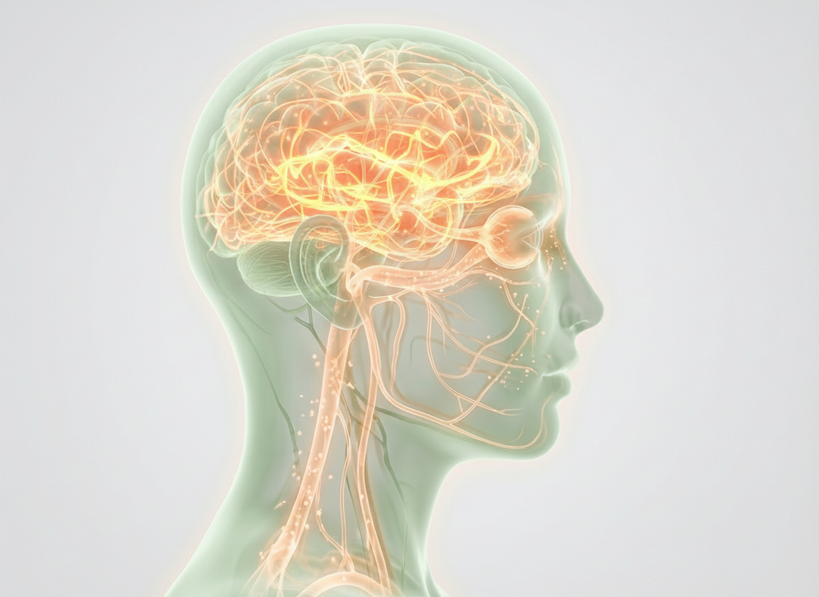
Most people who take omega-3 supplements do so, at least partly, because they've heard DHA supports brain health. That belief is scientifically reasonable — DHA is the dominant omega-3 in neuronal membranes, making up approximately 97% of the brain's total omega-3 content. The adult brain is roughly 60% fat, and DHA is a primary structural component of neuronal and photoreceptor membranes.
What is less widely understood is that getting DHA into the brain is a selective, transporter-mediated process — not a passive diffusion problem that more DHA in the blood simply solves. The same is true for the retina.
Nutrascience Neuro+ LPC is formulated specifically around this distinction. This article explains the mechanism, the evidence behind it, and how the formulation addresses both targets.
The barrier problem: why most DHA doesn't reach the brain or retina
The brain and retina are among the most metabolically demanding tissues in the body. They are also among the most carefully protected — shielded by highly selective barriers that control what enters from the bloodstream.
The blood-brain barrier (BBB) is formed by specialised endothelial cells lining brain capillaries. These cells are joined by tight junctions that prevent most substances from passing between them. Nutrient entry depends on specific transporter proteins embedded in the endothelial cell membrane. The blood-retinal barrier (BRB) is structurally distinct but functionally analogous: it protects the photoreceptor environment from unregulated systemic exposure using the same class of transporter proteins.
For DHA, both barriers share a critical gatekeeper: MFSD2A — Major Facilitator Superfamily Domain Containing 2A.
MFSD2A: the molecular gatekeeper
The identification of MFSD2A as the primary DHA transporter at the blood-brain barrier came from a landmark 2014 study published in Nature by Nguyen LN, Ma D, Shui G, et al. at Duke-NUS Medical School, Singapore, led by senior author David L. Silver. The researchers demonstrated that MFSD2A is expressed in the endothelium of the BBB and functions as a sodium-dependent lysophosphatidylcholine (LPC) transporter. Mice with the MFSD2A gene knocked out developed microcephaly and had dramatically reduced brain DHA levels — despite normal circulating DHA. The transporter, not the dose, was the limiting factor.
The critical mechanistic detail: MFSD2A recognises and transports DHA only when it is bound in the lysophosphatidylcholine (LPC) molecular form. DHA delivered in triglyceride or ethyl ester form — the molecular structure of standard fish oil and most algal DHA supplements — does not use this pathway efficiently.
A subsequent study by Alakbarzade V, Hameed A, Quek DQ, et al. (2015), published in Nature Genetics, confirmed that a partially inactivating mutation in the MFSD2A gene causes a non-lethal microcephaly syndrome in humans — establishing clinical relevance beyond mouse models.
Chan JP, Wong BH, Chin CF, et al. (2018), published in PLOS Biology, further characterised MFSD2A's role in brain development, demonstrating that LPC-DHA transported by MFSD2A acts as a physiological regulator of membrane phospholipid composition during a critical developmental window — identifying a feedback loop on lipogenesis that extends the functional significance of the transporter beyond simple DHA delivery.
The retinal case — why screen exposure makes this relevant now

For most of human history, the primary environmental stressor on photoreceptor cells was direct sunlight — a genuine concern, but one that most people limit through behaviour and hours of darkness.
The modern stressor is different. Sustained close-range screen exposure for 8–12 hours daily places continuous metabolic demand on photoreceptor cells. High-energy visible (HEV) light from screens does not establish the same phototoxicity risk as UV light at normal exposure levels. The mechanism is metabolic: sustained high-demand operation of photoreceptors, continuous renewal of outer segment membranes, and the oxidative load generated in the process.
Published research on retinal DHA (Bazan NG, 2009, Journal of Lipid Research) established the role of DHA in photoreceptor membrane function and the generation of docosanoids — DHA-derived signalling molecules including neuroprotectin D1 (NPD1) — that regulate cellular homeostasis and neuroprotective responses in the retina. DHA availability via the MFSD2A pathway directly supports the membrane quality that photoreceptor cells require to function at sustained metabolic demand.
For urban professionals with sustained screen exposure, the retina's DHA demand is quantitatively higher than it was for previous generations. This makes LPC-form DHA supply a more relevant variable than it would have been historically.
The same transporter operates at the retina
The retina contains photoreceptor cells — the rods and cones responsible for light detection — with some of the highest DHA concentrations in the body. These cells are among the most metabolically active in the human system, continuously consuming oxygen, generating oxidative byproducts, and renewing their outer segment membranes on a daily cycle. They depend on a continuous, adequate supply of DHA for membrane maintenance, signal transduction, and cellular signalling.
Critically, the blood-retinal barrier uses the same MFSD2A transporter as the blood-brain barrier. This was established by Wong BH, Chan JP, Cazenave-Gassiot A, et al. (2016), published in Journal of Biological Chemistry — identifying MFSD2A as operative at the retinal capillary endothelium and required for photoreceptor DHA accretion.
The practical implication: the molecular form requirement for DHA brain delivery applies equally to retinal delivery. Brain support and retinal support are not two different benefits of the same supplement. They are two applications of the same underlying biological mechanism — and both require LPC-form DHA to function efficiently.
What happens to DHA that isn't in LPC form
Standard fish oil and most algal DHA supplements deliver DHA in triglyceride (TAG) form. Krill oil delivers a portion of its DHA in phosphatidylcholine (PC) form, which is an improvement over triglyceride — but PC-DHA is hydrolysed by pancreatic phospholipase A2 during digestion, releasing free DHA that is then repackaged into triglycerides and circulated in chylomicrons.
Lagarde M, Bernoud N, Brossard N, et al. (2001), published in Journal of Molecular Neuroscience, established through radiolabelled DHA tracking that LPC-form DHA is the preferred carrier form across the blood-brain barrier, with significantly higher transfer efficiency than unesterified DHA through an in vitro BBB model.
Sugasini D, Thomas R, Yalagala PCR, et al. (2017), published in Scientific Reports, provided a direct dietary comparison: oral administration of LPC-DHA (40 mg DHA/kg) for 30 days in adult mice increased brain DHA content by more than 2-fold. The same amount of free DHA did not increase brain DHA — but did increase DHA in adipose tissue and heart. The study also reported improvements in spatial learning and memory with LPC-DHA that were absent in the free DHA group.
A note on the evidence: A 2025 replication study (Nutrients, Yalagala et al.) attempted to reproduce the Sugasini 2017 findings under tightly controlled conditions and reported no significant brain DHA increase with LPC-DHA at higher doses over a longer duration. This remains an active area of scientific discussion. The MFSD2A transport mechanism is established at Tier 1–2 evidence (genetic knockout studies, human mutation data, structural characterisation); the magnitude of brain DHA enrichment from dietary LPC-DHA in adult mammals is at a lower evidence tier and subject to ongoing investigation. We are transparent about this distinction rather than selectively citing favourable results.
The form problem, not the dose problem
The distinction matters because it reframes what "better omega-3 supplementation" means.
If the limiting factor were dose, the solution would be straightforward: take more fish oil. But if the limiting factor is molecular form — that MFSD2A requires LPC-bound DHA and most supplements deliver TAG-bound DHA — then increasing the dose of the wrong form does not address the bottleneck. It enriches peripheral tissues (adipose, heart, liver) without proportionally enriching brain or retinal tissue.
This is not a theoretical concern. The MFSD2A knockout data demonstrates that animals with adequate circulating DHA but compromised LPC transport develop microcephaly and severe brain DHA deficiency. The transporter is rate-limiting in a way that plasma DHA concentration alone cannot compensate for.
How Nutrascience Neuro+ LPC addresses both targets

Nutrascience Neuro+ LPC is formulated around two complementary components, both from Aker BioMarine.
Lysoveta® — LPC-bound omega-3s for the MFSD2A pathway
Lysoveta® is an LPC-enriched krill phospholipid complex developed by Aker BioMarine, a Norwegian marine biotechnology company. It was introduced in November 2020 as the world's first commercially available LPC-EPA/DHA ingredient, backed by 40 patents across 16 countries, and received FDA New Dietary Ingredient (NDI) notification status in 2023.
Lysoveta delivers omega-3 fatty acids — primarily DHA, with EPA also present — specifically in the LPC molecular form that MFSD2A recognises. The krill source is MSC-certified and harvested from Antarctic waters under regulated sustainable harvesting protocols.
Per serving (2 capsules), the Lysoveta® component in Neuro+ LPC provides:
- 54 mg LPC-bound omega-3s — the form that accesses the MFSD2A pathway
- 80 mg total phospholipids — structural support for cell membranes
- 60 mg combined EPA + DHA + DPA from krill
The LPC content is the functionally significant fraction. This is a form selection, not a dose maximisation.
On the choline contribution: The lysophosphatidylcholine backbone that carries LPC-DHA also carries choline — a precursor to acetylcholine, the neurotransmitter involved in memory encoding, muscle control, and nervous system signalling. This choline is intrinsic to the phospholipid structure, not a separately added ingredient. It is a downstream consequence of selecting the correct molecular form, not a primary rationale.
Revervia® — algal DHA for systemic support
Brain-targeted delivery addresses one pathway. The rest of the body — cardiovascular system, retina via systemic circulation, joints, and cells — still requires omega-3 support through standard absorption pathways.
Revervia® is a pure algal DHA oil derived from land-cultivated Schizochytrium sp. microalgae, also from Aker BioMarine. It delivers DHA in conventional triglyceride-form absorption, entering systemic circulation and distributing broadly.
Per serving, the Revervia® component provides approximately 244 mg DHA (calculated from COA: 61g/100g DHA × 400mg Revervia per serving), alongside EPA for systemic omega-3 status.
The case for algal DHA for the systemic component:
Sustainability. Revervia® is land-cultivated — it does not require ocean harvesting. Algae are the original biological source of omega-3s in the marine food chain; fish accumulate DHA by consuming them. Going directly to the source eliminates the intermediary.
Contaminant profile. Large fish bioaccumulate heavy metals, PCBs, and other environmental contaminants through the food chain. Land-cultivated microalgae carry none of this burden. For a supplement intended for long-term daily use, contaminant profile matters.
DHA purity. Revervia® delivers a high, consistent DHA concentration without the variable EPA ratios of fish-derived oils, which can complicate targeted DHA supplementation.
The combination addresses both components of the problem: LPC-pathway delivery for the brain and retina, and systemic omega-3 status for everything else.
How Neuro+ LPC compares
| Feature | Standard Fish Oil | Basic Krill Oil | Neuro+ LPC |
|---|---|---|---|
| DHA form for brain/retinal delivery | Triglyceride (limited MFSD2A use) | Phospholipid (partial) | LPC via Lysoveta® — MFSD2A-targeted |
| LPC-specific omega-3s | None | Minimal | 54 mg per serving |
| Total phospholipids per serving | None | Variable | 80 mg |
| Systemic DHA per serving | Variable (often 100–200 mg) | Typically low (50–80 mg) | ~244 mg (Revervia® algal DHA) |
| DHA source | Ocean fish | Ocean krill | Krill (LPC) + Land-cultivated algae (TG) |
| Sustainability | Ocean-dependent | Some MSC options | MSC-certified krill + land-cultivated algae |
| FDA NDI status | N/A | Varies | Yes — Lysoveta® (2023) |
| Contaminant risk | Higher (bioaccumulation) | Lower | Minimal (algal source + certified krill) |
| Vegetarian component | No | No | Revervia® DHA is fully vegetarian |
| Choline from phospholipid complex | No | Some | Yes — from phospholipid complex |
Brain and retinal health: what the research context shows

On MFSD2A and LPC transport (Tier 1–2 evidence):
The identification of MFSD2A as the primary brain DHA transporter, its substrate specificity for LPC-form fatty acids, and its clinical significance (human microcephaly mutations) represent well-established mechanistic science. Genetic, structural, and clinical convergence supports this at a high evidence tier.
On brain DHA enrichment from dietary LPC-DHA (mixed evidence):
Animal studies by Sugasini et al. (2017) reported substantial brain DHA enrichment from dietary LPC-DHA. This finding has not been consistently replicated. The mechanism is plausible and supported by transporter biology, but the magnitude of effect in adult humans from supplemental doses remains to be established by well-powered human RCTs. We present the animal data as directionally relevant, not as established clinical proof.
On retinal DHA and screen exposure:
The role of DHA in photoreceptor membrane function is well-characterised. The metabolic demand hypothesis for modern screen exposure is biologically coherent but not yet the subject of dedicated clinical trials. Observational associations between omega-3 status and retinal health exist; causation requires prospective intervention data.
On population-level associations (Tier 3 evidence):
Epidemiological studies observe associations between omega-3 status and cognitive trajectory over time. These are directionally consistent but cannot establish causation in observational design.
This content is educational. Nutrascience Neuro+ LPC is a nutritional supplement, not a treatment for macular degeneration, Alzheimer's disease, or any other condition. For anyone with diagnosed neurological or ocular conditions, the appropriate starting point is a healthcare professional.
Key references
- Nguyen LN, Ma D, Shui G, et al. Mfsd2a is a transporter for the essential omega-3 fatty acid docosahexaenoic acid. Nature. 2014;509(7501):503–506.
- Alakbarzade V, Hameed A, Quek DQ, et al. A partially inactivating mutation in the sodium-dependent lysophosphatidylcholine transporter MFSD2A causes a non-lethal microcephaly syndrome. Nature Genetics. 2015;47(7):814–817.
- Chan JP, Wong BH, Chin CF, et al. The lysolipid transporter Mfsd2a regulates lipogenesis in the developing brain. PLOS Biology. 2018;16(8):e2006443.
- Wong BH, Chan JP, Cazenave-Gassiot A, et al. Mfsd2a is a transporter for the essential omega-3 fatty acid docosahexaenoic acid in the retina. Journal of Biological Chemistry. 2016;291(20):10501–10514.
- Lagarde M, Bernoud N, Brossard N, et al. Lysophosphatidylcholine as a preferred carrier form of docosahexaenoic acid to the brain. Journal of Molecular Neuroscience. 2001;16(2–3):201–204.
- Sugasini D, Thomas R, Yalagala PCR, et al. Dietary docosahexaenoic acid (DHA) as lysophosphatidylcholine, but not as free acid, enriches brain DHA and improves memory in adult mice. Scientific Reports. 2017;7:11263.
- Bazan NG. Neuroprotectin D1-mediated anti-inflammatory and survival signalling in stroke, retinal degenerations, and Alzheimer's disease. Journal of Lipid Research. 2009;50 Suppl:S400–5.
Where can I get Neuro+ LPC?
Free shipping to SG, MY, HK & Bangkok , on orders above MYR200.00. Order Nutrascience Neuro+ LPC →
Full biological timeline — what's happening and when
The mechanism here is structural incorporation, not acute pharmacological effect. Understanding what is happening biologically at each stage is the correct frame for evaluating whether the supplement is working.
Weeks 1–4: Establishing bioavailability
With consistent daily dosing, circulating LPC-DHA levels rise. Blood plasma LPC-DHA concentrations begin to increase — this is the molecular form that the MFSD2A transporter at the blood-brain and blood-retinal barriers recognises.
What's happening physiologically: The transporter now has its substrate available. It can begin moving LPC-DHA across both barriers into neural and retinal tissue. Nothing is perceptible at this stage. This is the foundation phase.
Why stopping here is a mistake. Steady-state plasma LPC-DHA levels are still building. Stopping supplementation before steady state is established means the mechanism never had a fair opportunity to operate.
Weeks 4–8: Active membrane incorporation — brain and retina
Brain phospholipid turnover is a continuous biological process. Neurons regularly replace and remodel their membrane phospholipids. With elevated plasma LPC-DHA available, DHA begins incorporating into neuronal membranes — particularly in regions with high metabolic demand.
The same process is occurring in the retina. Photoreceptor membrane turnover is among the fastest in the body — the outer segments of rod and cone cells are continually renewed, shedding and replacing membrane material on a cycle of days. With LPC-DHA available via the MFSD2A pathway at the blood-retinal barrier, this renewal process draws on a higher-quality substrate.
The hippocampus (memory), prefrontal cortex (sustained attention and executive function), and retinal photoreceptors are all incorporating DHA in this window.
What some people notice: subtle improvements in mental clarity or reduced eye fatigue during sustained screen work. Not universal. Not guaranteed. But these reports align with the biological timeline of active DHA incorporation into both neural and retinal membrane structures.
Weeks 8–12 and beyond: Stabilisation at a higher baseline
Tissue DHA concentrations stabilise. Brain and retinal membranes now operate at higher DHA density — supporting membrane fluidity, synaptic signalling efficiency, and cellular resilience to oxidative stress in metabolically demanding tissues.
This is not a dramatic threshold. There is no point at which you suddenly "feel" your brain DHA levels have risen. Structural support works below the threshold of perception — which is precisely what distinguishes it from stimulants and acute interventions.
The analogy: you don't feel your muscles synthesising protein after training. Structural support operates on biological timelines and biological scales. Nutrascience Neuro+ LPC is no different.
The significance of consistent daily dosing
DHA incorporation into brain and retinal membranes is a slow, continuous biological process. The rate of incorporation depends on plasma DHA availability. The rate of loss follows a similar slow curve when supplementation stops.
Evidence on DHA washout kinetics indicates that plasma DHA levels begin declining within days of stopping supplementation, and tissue DHA levels decline more gradually over weeks. Consistent daily dosing maintains the elevated plasma pool the transport mechanism draws from.